Leadership in Biotech

Tag: open-source

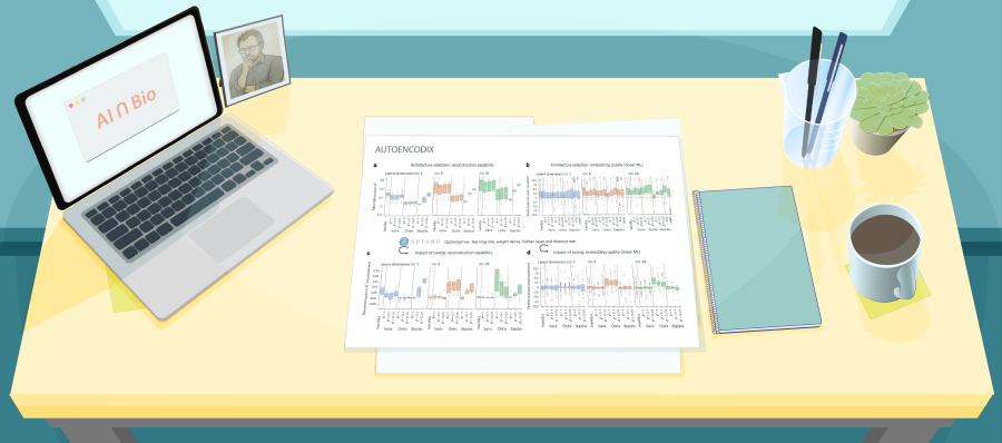
Illustration of a desk with a figure from a recent paper about AUTOENCODIX

Interpretable Latent Spaces, Messy Biology: What AUTOENCODIX Teaches Us About Autoencoders in the Wild

This week in AI ∩ Bio I dug into AUTOENCODIX, an open-source framework that stress-tests autoencoders (AEs) on real multi-omics data.

The punchline: no single architecture wins, reconstruction scores can mislead, and “interpretable” latent spaces inherit every bias baked into our ontologies. This paper provides exactly the kind of clarity we need to effectively apply these models in diagnostics and biomarker discovery.

AUTOENCODIX is a new open-source framework that tries to bring order to the AEs chaos in multi-omics, allowing the user to test multiple AEs through the same pipeline, then compare not just loss curves but how useful the learned embeddings actually are for biology and prognosis. (AEs explained in carousel)

Scientifically, a few themes stood out:

  1. they show how tuning β in VAEs affects performance; low β favors reconstruction; high β imposes compact, disentangled latent spaces.
  2. across TCGA and single-cell cortex data, no AE architecture consistently outperforms others. Good reconstruction doesn’t guarantee useful embeddings. Ontix, the biologically structured AE, wires decoder layers to known pathways or chromosomes, making latent dimensions interpretable. But robustness varies and depends on learning rate; and the results hint at artifactual learning (see comments).

Diagnostics-leadership perspective

This paper is a reminder to separate infrastructure from insight.

AUTOENCODIX is essentially AE infrastructure: it standardizes data handling, model training, and evaluation so you can ask disciplined questions instead of chasing whichever architecture is trending.

The results also challenge the reflex to equate fancier models with better clinical value: PCA remains a very strong baseline, and ontology-based models only shine when the chosen ontology matches the question and is treated carefully as a potential source of bias, not ground truth.

For leaders deciding where to invest, the take-home is: fund frameworks that make comparisons fair and reproducible, and judge models by task-relevant endpoints and robustness across cohorts—not by reconstruction loss or aesthetic latent plots.
For early-career scientists

There’s a quieter lesson here about how to work with powerful tools without giving up your scientific spine.

The authors don’t present a magical autoencoder that “solves” multi-omics; instead, they map trade-offs, show when tuning helps and when it doesn’t, and surface uncomfortable findings like decreased robustness after hyperparameter optimization for ontology-based VAEs.

If you’re building a career in computational or experimental biology, papers like this are an invitation to open the hood: run the benchmarks, break the assumptions, test models on tasks you actually care about, and treat interpretability as something you design and stress-test—not something you assume.

Skip to PDF content
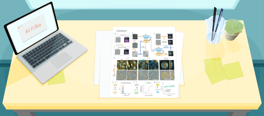
Illustration of a desk with figures from a paper on Cytoland

The Model Isn’t the Magic: How Cytoland shows that domain expertise—not just deep learning—is what makes AI in biology work.

This week’s AI ∩ Bio: Reading the Revolution series covers Cytoland, a collection of models for robust virtual staining of landmark organelles across diverse imaging parameters, cell states and types.

The core idea of Cytoland

Cytoland introduces a set of deep learning models for virtual staining—predicting fluorescent labels of key organelles (nuclei, membranes) from label-free microscopy data like quantitative phase images (QPI). These models overcome current limits in generalization, robustness, and data efficiency, offering practical tools for label-free live-cell phenotyping across multiple cell types, imaging setups, and biological contexts.

Basically, the paper introduces a methodology to use AI to predict what a fluorescent image would look like—based only on label-free data (like phase contrast or brightfield). Think of it as teaching the model to “see” the nucleus or membrane without physically tagging them.

Why this matters

Virtual staining isn’t just a cool trick—it can fundamentally shift how we do live-cell imaging, avoiding time and engineering resources to make fluorescent labels for every experiment. And crucially, Cytoland models are open-source, tested on real-world problems, and robust enough to use across microscopes, labs, and cell systems.

In the AI ∩ Bio landscape

This paper is a landmark example of precision engineering, not algorithmic revolution. It does what so many papers fail to do: combine deep learning, physics, and real experimental constraints into a system that’s usable and robust.

You don’t get this kind of robustness without deep domain expertise. You have to understand how microscopes work, what makes a label “missing” vs. “invisible,” and how to design models that hold up in the real world. The magic happens not in the model itself, but at the intersection of thoughtful AI design, careful data collection, and biological insight.

For early-career scientists

The breakthrough isn’t just in the model—it’s in:

  • Asking biologically meaningful questions
  • Collecting the right data
  • Designing AI that respects the physics and messiness of real experiments

You don’t need to invent new algorithms to do impactful AI ∩ Bio work.

What matters more is:

  • Understanding what matters biologically (e.g., membranes over time, infection states, tissue development)
  • Designing training and validation that respects experimental complexity
  • Building models that generalize to the messiness of real world data

Learn how to ask scientific questions that AI can help answer, and how to design datasets and metrics that hold models accountable to real biological use.

Powered by WordPress & Theme by Anders Norén