Leadership in Biotech

Author: Kristin Gleitsman Page 4 of 7

Scientist. Research and Development Leader. Collaborative Problem Solver.
Passionate about building belonging, so that together we can accomplish important, hard things.

Illustration of a desk with a figure from a recent paper about AI interpretability in biology

What Are Genomic Transformers Actually Learning?

This week’s AI ∩ Bio: Reading the Revolution post explores a new paper tackling one of the biggest challenges in AI for biology: interpretability.

Specifically, how can we tell what genomic transformer models are learning and whether those internal patterns map to real biological concepts?

The study proposes a systematic framework to analyze attention heads in models like DNABERT and scGPT. It reveals that many attention heads naturally align with key biological features—such as transcription start sites, sequence motifs (e.g., TATAAA), GC content, conservation scores, and Gene Ontology categories like “neuron projection” or “mitochondrial membrane.” This interpretability emerges even during self-supervised pre-training and strengthens with task-specific fine-tuning.

Key Findings

  • Attention heads develop strong correlations with biologically meaningful features.
  • These heads tend to be more important for predictive performance, as shown by ablation experiments.
  • Interpretability is shaped by tokenization strategy (e.g., overlapping vs. non-overlapping k-mers).
  • GPT-4 was used to generate descriptive summaries of head activity (e.g., “TATA box detector”)—but these require validation, as they can miss label-specific nuance.

Limitations

  • Correlation ≠ causation: attention may highlight a feature without depending on it for prediction.
  • Current analyses treat attention heads in isolation—without clustering or organizing them into higher-level “concept modules.”
  • GPT-4 summaries, while useful, may overgeneralize or obscure context-dependent patterns.

Why This Matters

This work moves the field closer to a compelling vision: transformer models that don’t just output predictions, but expose interpretable biological patterns that scientists can name, reason about, and test. Much like ConcepPath in digital pathology that we covered earlier in this series, this paper hints at a future where AI supports concept-level ‘reasoning,’ not just classification.

For Early-Career Scientists

Interpretability isn’t just about trust, it’s also critical for scientific translation. These models may surface patterns that align with biology, but the real opportunity is in bridging those signals to concepts we recognize, can reason about, and critically, can test experimentally. That’s how interpretability becomes a tool for discovery, not because the model “knows” biology, but because we can now ask better questions about what it might be pointing to.

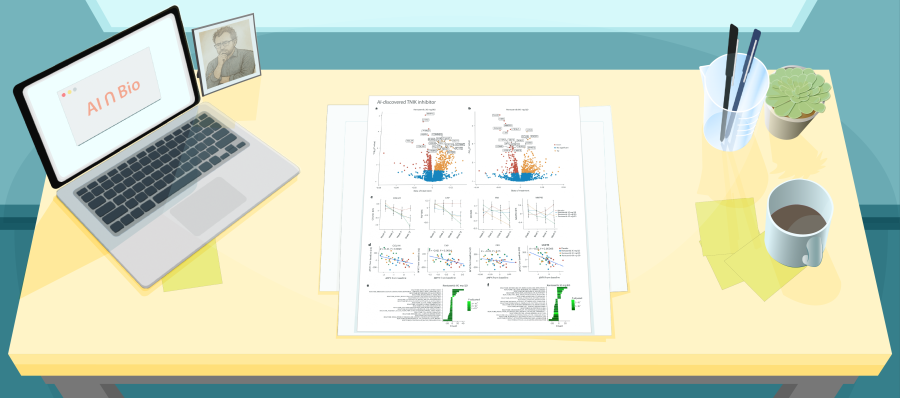
Illustration of a desk with a figure from a recent paper about Deep-learning-based gene perturbation

When Complexity Misleads

In this week’s AI ∩ Bio paper, we look at a multi-dataset benchmark where deep learning models trained to predict single-cell perturbation outcomes did not outperform two deliberately simple baselines: “assume nothing changes” and “predict the additive combination of single perturbations.” Performance improved when models were reduced to just their internal embeddings and a linear decoder—no deep layers, just a direct projection from embedding space to gene expression. In other words, the models had learned biologically meaningful representations during training, but using those representations to make predictions through a complex decoder often made things worse.

This result may not surprise those familiar with traditional machine learning approaches. As the bias–variance tradeoff reminds us: when signal is weak and noise is high, simpler models often generalize better and overfit less, leading them to outperform more complex alternatives.

This study revealed both conditions:

  • The signal from genetic perturbations is often modest or additive.
  • The noise in single-cell gene expression is high, due to dropout, amplification bias, and biological variability.

If your model must detect subtle shifts in expression, but your measurement system is saturated with noise, then even an optimal model will struggle to generate meaningful predictions. And a large model may do worse, because it fits spurious correlations that simpler models ignore.

Before adding more architecture, it’s worth asking:

  • Have we clearly defined our outcome?
  • Do we understand the system we are working with?
  • Can our data realistically support the question we’re asking?

Otherwise, we risk building a computational Rube Goldberg machine: impressive, fragile, and ultimately unclear.

Illustration of a desk with a figure from a recent paper about Rentosertib

Hype, Hurdles, and Hepatotoxicity: A Bold Step for AI-Designed Drugs, But Still Miles to Go

This week’s AI ∩ Bio: Reading the Revolution paper spotlights the first randomized Phase 2a trial of a drug discovered using generative AI.

Summary and Context

The trial investigates rentosertib, a small-molecule inhibitor of TNIK (Traf2- and Nck-interacting kinase), developed end-to-end on Insilico’s AI platform. Notably, the system identified both the target and compound de novo—compressing two traditionally distinct phases of drug discovery into a unified AI-led workflow.

The study enrolled patients with idiopathic pulmonary fibrosis (IPF)—a progressive, fatal lung disease with no cure. This trial sets a bold precedent: Can a molecule born of algorithms succeed in one of medicine’s most unforgiving indications?

Scientific Insights & Critical Observations

What’s Promising

  • AI-led discovery pipeline: TNIK identified and rentosertib designed via Insilico’s generative model.
  • Accelerated early development: Reached Phase 2a enrollment in under 30 months from target nomination.
  • Biomarker engagement: Downregulation of fibrosis-linked proteins (e.g., COL1A1, MMP10, FAP) with exploratory ties to lung function improvement.

What’s Concerning

  • Overinterpretation of early signals: The +98.4 mL FVC gain in the 60 mg arm was non-significant, with one-third of patients missing spirometry at Week 12.
  • Safety flags: Hepatotoxicity led to multiple discontinuations, particularly in patients co-treated with nintedanib—a standard-of-care antifibrotic—with an unexplored drug–drug interaction risk.
  • Pharmacokinetics: High interpatient variability and disproportionate exposure increases raise questions about dose optimization.
  • Generalizability: All 71 patients were Asian and enrolled in China, limiting extrapolation across diverse IPF populations.
  • Narrative overreach: The AI platform is being validated rhetorically more than pharmacologically—so far.

A Useful Comparison

To contextualize this AI-driven advance, consider nintedanib—a first-in-class oral tyrosine kinase inhibitor targeting PDGFR, FGFR, and VEGFR. It was the first drug to significantly slow lung function decline in IPF in large, well-controlled trials, establishing antifibrotic therapy as a treatment paradigm.

FeatureRentosertib (AI, Phase 2a, 2025)Nintedanib (Traditional, Phase 2 results in 2011- below; Approved c2014)
Discovery speed~30 months to Phase 2a~10+ years
Target noveltyFirst-in-class (TNIK)Known pro-fibrotic RTKs
Phase II trial size71 patients432 patients
Efficacy signalWeak, non-significantStatistically significant
Safety profileHepatotoxicity, DDI with nintedanibPredictable, tolerable
Trial population diversitySingle-country, homogeneous cohortGlobal, multi-ethnic
Biomarker integrationProteomics exploredFocused on validated clinical endpoints

Takeaway for Early-Career Scientists

This is a milestone in ambition, not yet in outcome.
Rentosertib shows what generative AI can compress in timeline—but not what it can yet deliver in therapeutic benefit. The real test isn’t how a drug is designed, but how it performs in humans.

So ask yourself: If this same molecule came from a traditional screen, would we be touting it—or shelving it?

Generative AI may change how we create drugs, but it doesn’t change what success looks like.

Illustration of a desk with a figure from a recent paper about AlphaFold3 Evaluations

Testing the Physics Beneath the Predictions Beyond RMSD: What AlphaFold3 Really Understands

This week’s paper is personal for me.

Dan Herschlag was my postdoc advisor. Dan taught me how to think mechanistically, how to test assumptions, and how to pursue scientific clarity with unflinching rigor.

That legacy is all over this paper. And is incredibly needed in this era of AI hype where data volume often sidelines careful model-driven science.

Herschlag et al. show that while AlphaFold3 predicts protein structures with backbone-level structural precision, it struggles to capture the physical rules that govern biological function.

But this paper isn’t a takedown: it’s a roadmap for how to go further.

What They Did

Rather than rely on RMSD alone, the team evaluated AlphaFold2 and AlphaFold3 against:

  • Energetic rules: bond torsions, hydrogen bonds, and van der Waals contacts
  • Experimental ensembles: from multi-temperature crystallography
  • Model confidence: comparing pLDDT to physical plausibility

What They Found

  • ~30% of side-chain interactions deviated from experimental observations, often with incorrect partners or implausible geometries
  • High-confidence predictions (>90 pLDDT) still showed strained or physically invalid conformations
  • Energetically-favorable conformations could increase RMSD and be penalized by the model
  • AlphaFold3 missed ~85% of conformational variability seen in real experimental ensembles

Key Insight: Physics ≠ Proximity

AlphaFold can recapitulate Ramachandran and Lennard-Jones-like patterns. But that doesn’t mean it understands physical constraints.
To improve these tools, we need evaluation metrics grounded in molecular energetics, not just geometry.

Takeaway for Scientists

This paper is a reminder that progress requires more than prettier predictions. It demands models that reflect the physics that drive biology.

Illustration of a desk with a figure from a recent paper about binding affinity predictions

Beyond Binding: Rethinking Drug Design in the Age of AI and Structural Biology

In molecular design, we often prioritize what’s measurable over what’s meaningful.

For decades, binding affinity has served as a cornerstone of early-stage drug discovery, not because it captures biological function in full, but because it’s one of the few properties we can quantify systematically and optimize across large libraries.

Now, as AI models generate binders faster than we can validate them, we must ask: What exactly are we optimizing for? And what datasets are we training on?

What we know:

  • Strong binding doesn’t guarantee efficacy
  • Residence time and conformational flexibility can matter more than affinity
  • Cellular context — target expression, pathway crosstalk, and off-target interactions — often dictates outcome in clinical applications

Yet much of the public data — and many AI training sets — still orbit around Kd, IC₅₀, and docking scores. These are abundant and easy to label, but they capture only a narrow slice of pharmacological reality (and we’re not even accounting for the fact that these measurements are highly dependent on the specific conditions- buffer, temperature, etc).

If we train models on what’s easy to measure, we shouldn’t be surprised when they generate molecules that impress in silico — and disappoint in vivo.

The problem isn’t that binding doesn’t matter. It does. The problem is that binding isn’t biology.

Toward More Meaningful Models

To do better, we’ll need to:

  • Incorporate multi-parametric data: kinetics, permeability, metabolism, toxicity, immune activation
  • Train models to include mechanism and uncertainty, not just affinity
  • Elevate datasets that link structure to systems, not just structure to scores

The best work ahead won’t just generate molecules; it will surface better models about how they work, and where they fail in the journey from discovery to clinic.

Illustration of a desk with a figure from a recent paper about Graphinity

Investigating the volume and diversity of data needed for generalizable antibody–antigen ΔΔG prediction

This week’s AI ∩ Bio: Reading the Revolution paper isn’t just a new model or a shiny dataset. It’s a reality check that cuts through superficial benchmark performance to ask harder questions about AI for antibody design.

While many models report high accuracy on test sets that closely resemble their training data, Hummer et al. go further, testing how models perform when challenged with unseen antibody–antigen complexes. This approach led to a rigorous investigation of why models fail, and what it would take to make ΔΔG prediction truly generalizable.

They introduce Graphinity, a graph neural network trained to predict changes in antibody–antigen binding affinity (ΔΔG). They rigorously stress-test the system: varying sequence identity cutoffs, benchmarking against other architectures, simulating noisy labels, and even filtering mutations by evolutionary plausibility.

The findings are clear and sobering: it’s not the model that’s the bottleneck. It’s the data: data size, data diversity, and data alignment with real-world biological utility.

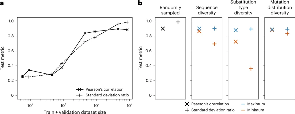
Graphs showing considerations for experimental ΔΔG dataset generation, with respect to ML predictiveness

a,b, Model performance with varying training plus validation dataset size

Scientific Insight

The core contribution isn’t Graphinity’s design (though it performs well). It’s the authors’ commitment to not mistaking narrow wins for general insight. They show that a 0.9 Pearson correlation can be misleading if training and test data are too similar. That scaling dataset size doesn’t help if you’re mutating the same few complexes. And posit that no model—regardless of architecture—can learn to generalize if it’s never exposed to true diversity.

Crucially, they quantify the gap: at least 90,000 experimental ΔΔG values may be needed to reach robust generalization. And they begin to chart how we might get there—by prioritizing diverse substitutions, broad antigen coverage, realistic effect sizes, and evolutionarily grounded mutations—not just alanine scans.

To R&D leaders

This paper demonstrates what it looks like to take biological realism seriously. If you’re evaluating models for therapeutic antibody design, don’t just ask about top-line metrics. Ask how performance changes when similar complexes are removed from the test set. Dig deeply into what signal the model is learning and whether that signal will hold for the next antigen.

To early-career scientists

This paper helps us see what not fooling yourself looks like. It’s a reminder that model performance only matters when grounded in data that reflects the system’s true complexity. If you want to build models that generalize, start by designing experiments that enable that.

Boltz-2: How much can 3D structure really tell us about molecular binding energetics?

This week’s AI ∩ Bio: Reading the Revolution series covers Boltz-2, a new structural biology foundation model that exhibits strong performance for both structure and affinity prediction.

To put this work in context, let’s start with the classic protein modeling pipeline logic:
🧬 Sequence → 🧱 Structure → 🎯 Function

AlphaFold revolutionized the first step, grounded in the premise that function follows from structure. Boltz-2 puts that premise to the test. It starts at the middle of the pipeline — with the 3D structure of a protein–ligand complex — and asks: Can we predict binding affinity using only geometry?

Structure is signal

Boltz-2 is a deep learning model that predicts binding affinity directly from 3D geometry — no sequence, no docking scores, no molecular dynamics.
It learns by:

  • Using real 3D snapshots of protein–ligand complexes from experiments (via the PDBBind database) as “correct” examples
  • Comparing them to incorrect or nonbinding versions (decoys)
  • Teaching itself to distinguish between the two by assigning higher scores to the true binders — a method called contrastive learning
  • Viewing each complex from multiple angles and modeling how atoms interact using cross-attention between the ligand and protein

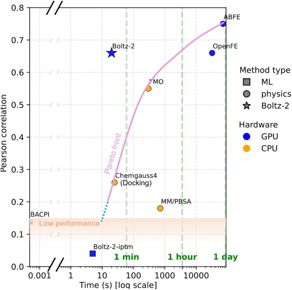
The result? Accuracy approaching Free Energy Perturbation (FEP) — a gold-standard physics-based method — at a fraction of the computational cost. So if you have the correct structure, you can get binding affinity. But that’s the tradeoff.

Boltz-2 doesn’t predict binding sites. It doesn’t model flexible loops or conformational dynamics. It assumes the structure is already known — and that it’s accurate. But we know that:

  • Crystallography can trap proteins in inactive states
  • Ligand poses may not reflect behavior in solution
  • Flexibility is collapsed into a single static frame

Still, Boltz-2 shows how much signal is embedded in structure — when that structure is right.


Reflection for Early-Career Scientists

What happens when you flip the framing? Instead of building up from sequence to structure to function, Boltz-2 works from the middle, assuming structure is known, and asking how far that alone can take you. As a result, Boltz-2 sharpens the boundary of what structure can predict — and what it can’t. In other words, Boltz-2 is a boundary marker: a way to measure what’s possible if geometry is complete and correct.

Graph of Pearson Correlations over time, showing Boltz-2 with a strong accuracy / speed trade-off for affinity prediction

Boltz-2 presents a strong accuracy / speed trade-off for affinity prediction.

Illustration of a desk with figures from a paper on Cytoland

The Model Isn’t the Magic: How Cytoland shows that domain expertise—not just deep learning—is what makes AI in biology work.

This week’s AI ∩ Bio: Reading the Revolution series covers Cytoland, a collection of models for robust virtual staining of landmark organelles across diverse imaging parameters, cell states and types.

The core idea of Cytoland

Cytoland introduces a set of deep learning models for virtual staining—predicting fluorescent labels of key organelles (nuclei, membranes) from label-free microscopy data like quantitative phase images (QPI). These models overcome current limits in generalization, robustness, and data efficiency, offering practical tools for label-free live-cell phenotyping across multiple cell types, imaging setups, and biological contexts.

Basically, the paper introduces a methodology to use AI to predict what a fluorescent image would look like—based only on label-free data (like phase contrast or brightfield). Think of it as teaching the model to “see” the nucleus or membrane without physically tagging them.

Why this matters

Virtual staining isn’t just a cool trick—it can fundamentally shift how we do live-cell imaging, avoiding time and engineering resources to make fluorescent labels for every experiment. And crucially, Cytoland models are open-source, tested on real-world problems, and robust enough to use across microscopes, labs, and cell systems.

In the AI ∩ Bio landscape

This paper is a landmark example of precision engineering, not algorithmic revolution. It does what so many papers fail to do: combine deep learning, physics, and real experimental constraints into a system that’s usable and robust.

You don’t get this kind of robustness without deep domain expertise. You have to understand how microscopes work, what makes a label “missing” vs. “invisible,” and how to design models that hold up in the real world. The magic happens not in the model itself, but at the intersection of thoughtful AI design, careful data collection, and biological insight.

For early-career scientists

The breakthrough isn’t just in the model—it’s in:

  • Asking biologically meaningful questions
  • Collecting the right data
  • Designing AI that respects the physics and messiness of real experiments

You don’t need to invent new algorithms to do impactful AI ∩ Bio work.

What matters more is:

  • Understanding what matters biologically (e.g., membranes over time, infection states, tissue development)
  • Designing training and validation that respects experimental complexity
  • Building models that generalize to the messiness of real world data

Learn how to ask scientific questions that AI can help answer, and how to design datasets and metrics that hold models accountable to real biological use.

Illustration of a desk with figures from a paper on ConcepPath

From Better Models to Better Questions: A Pathology AI Rethink

This week’s AI ∩ Bio: Reading the Revolution series covers ConcepPath, a new framework that blends expert-derived pathology concepts with deep learning to improve both accuracy and interpretability in histopathology image analysis. Instead of relying only on slide-level labels (e.g., “adenocarcinoma”), ConcepPath uses GPT-4 to extract detailed visual concepts from medical literature and aligns them with tissue regions using vision-language models trained on pathology image–text pairs, helping explain predictions in terms that reflect how diagnoses are taught, documented, and defended in clinical practice.

Scientific Approach

Most AI models for whole slide images (WSIs) use Multiple Instance Learning (MIL); they divide slides into patches, analyze each one, and then aggregate the predictions. This works for classification but offers little insight into why a decision was made. NOTE: more on the ‘standard’ approach in the carousel. ConcepPath adds a critical layer: concept alignment. First, GPT-4 infers visual pathology features from peer-reviewed literature—these become expert-informed concepts. The model then learns additional data-driven patterns directly from the images themselves, potentially novel features that improve prediction even if they lack clinical names. These concepts, both known and learned, are aligned with image features using CLIP-style models (Contrastive Language–Image Pretraining, or models that learn to match images and text that describe the same thing). The model then produces similarity maps showing which parts of the tissue match each concept, for example, highlighting keratin pearls in a region suggestive of squamous carcinoma.

These maps improve interpretability, but they do not replicate diagnostic reasoning. Still, this structured mapping makes model outputs more traceable and aligned with how pathologists evaluate slides.

A Step Forward in Trustworthy AI?

ConcepPath signals a strategic evolution in diagnostic AI—from black-box performance to structured, clinically-aligned transparency. While attention maps and saliency overlays are increasingly common, ConcepPath distinguishes itself by embedding domain knowledge into the model architecture itself. It doesn’t just show where the model looked—it tells us what features it saw.

For early-career researchers, this paper is a reminder that innovation doesn’t always come from better algorithms—it often comes from asking deeper questions. In this case, the authors asked: What would it take for AI to reason with the same visual vocabulary and decision cues as a pathologist? That mindset led to a model that’s both potentially better and more explainable. In computational medicine, it’s this pairing of technical skill and conceptual clarity that sets the stage for meaningful impact.

Illustration of a desk with figures from a paper on X-Atlas/Orion

X-Atlas/Orion: Your Model is Only as Good as Your Training Data

I hope you will forgive me this week, because this week’s paper isn’t an AI paper per se… it’s a methods and data generation paper, but one that sits squarely at the intersection of AI and biology. While much of the field is focused on building more powerful models, even the best architectures can’t reliably infer causality, dose effects, or subtle regulatory responses from observational data alone. Controlled perturbations are essential for training models that work towards explanation, not just correlation. 

In this preprint, the team at Xaira Therapeutics, a next-gen biotech building AI-native drug discovery tools, introduces FiCS Perturb-seq, a scalable, industrialized platform for dose-aware single-cell CRISPR. And the open source dataset they generated using this method, X-Atlas/Orion, is something truly remarkable.  

Scientific Insight

The core breakthrough here isn’t a novel discovery, it’s the industrialization of perturbation data generation. FiCS Perturb-seq standardizes the full pipeline: fixation, cryopreservation, FACS enrichment, automation, and high-density single-cell loading. This enables X-Atlas/Orion, a dataset of 8 million dual-sgRNA cells with deep, reproducible transcriptomic profiles—built not for exploration alone, but for training dose-aware, causal foundation models. A key analytic innovation: using sgRNA abundance as a proxy for knockdown strength, enabling dose-dependent transcriptional analyses that move beyond the binary logic of most Perturb-seq studies.

For R&D leaders, this paper is a great example for how you can scale experimental biology to meet the needs of machine learning. It’s not about a single step, it’s about asking the right questions about what data is really needed and designing the system to generate that data at scale: QC before sequencing, consistent cell handling, and deep coverage per perturbation. 

To early-career scientists: this paper highlights the importance of infrastructure for discovery. The authors didn’t invent fixation or dual guides—they made them scalable, automatable, and reliable enough to support next-generation modeling. If you’re working at the interface of wet lab and ML, remember: your model is only as good as your training data. The signal (i.e., what am I really measuring and what can it actually tell me), the structure, and the variability you build into your system will define what the model learns and what it misses.

Page 4 of 7

Powered by WordPress & Theme by Anders Norén